A woman should talk with her health care provider about her personal risk of getting breast cancer. She should ask questions about when to start and how often to be checked for the disease. These decisions, like many other medical decisions, should fit each woman’s particular needs.
Screening for cancer before there are symptoms is important. It can help doctors find and treat cancer early and treatment is more likely to be effective when cancer is found early. Your health care provider may suggest screening tests to check for breast cancer before any symptoms develop:
- Screening mammogram
- Clinical breast exam
- Breast self-exam
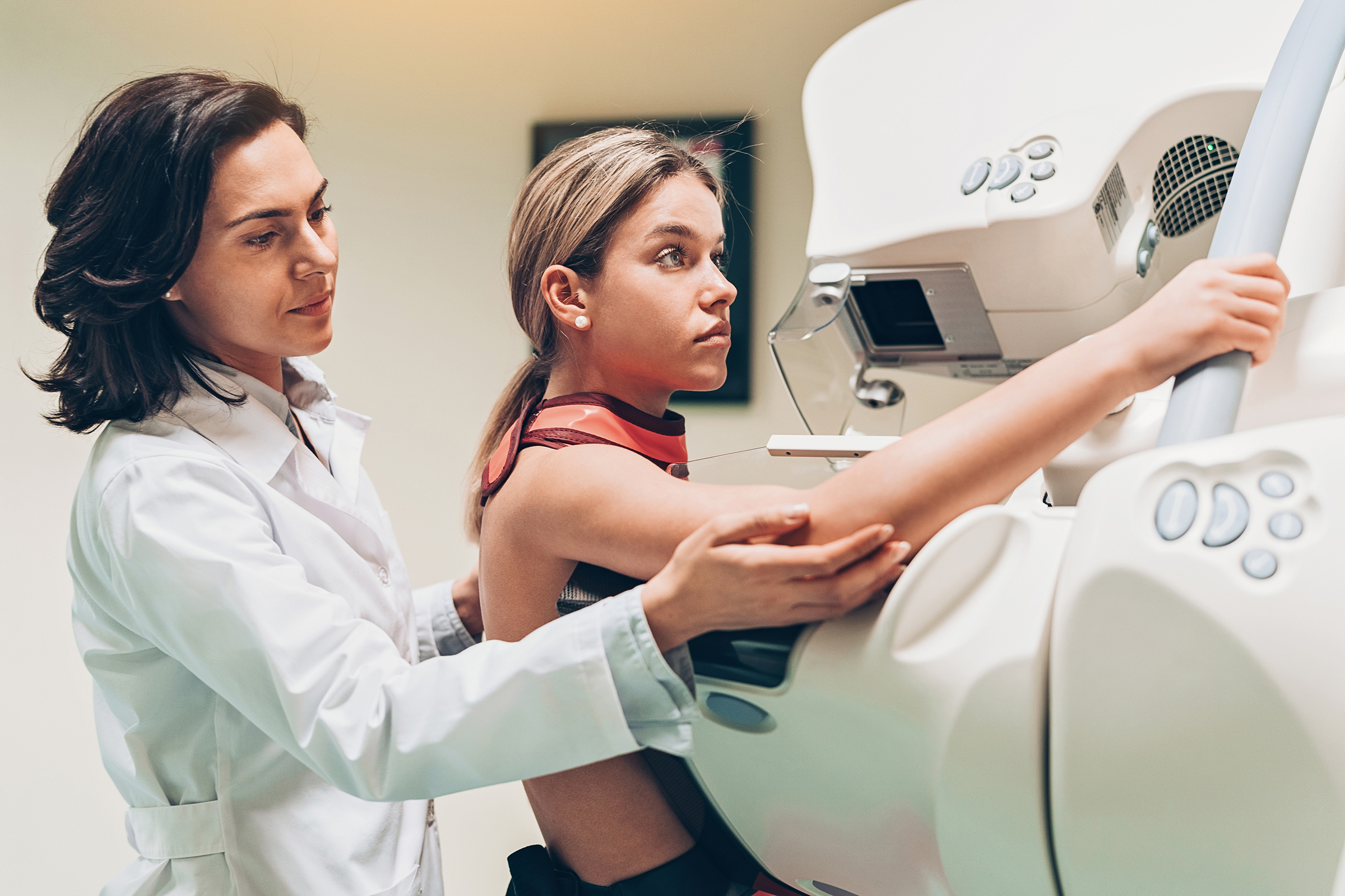
Screening Mammogram
A mammogram is used to find breast cancer early:
Women in their 40s and older should have mammograms every one to two years.
Screening mammograms can often show a breast lump before it can be felt. They also can show a cluster of very tiny specks of calcium. These specks are called microcalcifications. Lumps or specks can be signs of cancer.
If the physician sees an abnormal area on the mammogram, the woman may need more pictures taken. Also, the woman may need to have a biopsy. A biopsy is the only way to tell for sure if cancer is present.
Mammograms are the best tool doctors have to find breast cancer early. However, it is good for a woman to keep in mind that mammograms (as well as dental x-rays, and other routine x-rays) use very small doses of radiation.
Breast Exam
During a clinical breast exam, the health care provider feels the breasts while the woman is standing or sitting up and lying down. The woman may be asked to raise her arms over her head, let them hang by her sides, or press her hands against her hips.
The health care provider looks for differences between the breasts, including unusual differences in size or shape. The skin of each breast is checked for a rash, dimpling, or other abnormal signs. The nipples may be squeezed to see if fluid is present.
Using the pads of the fingers to feel for lumps, the health care provider checks the entire breast, the underarm, and the collarbone area, first on one side, then on the other. A lump is generally the size of a pea before anyone can feel it. The lymph nodes near the breast may be checked to see if they are swollen. A thorough clinical breast exam may take 10 minutes.
Breast Self-Exam
It’s important for women to perform monthly breast self-exams to check for any changes in their breasts. Keep in mind when you do this exam that each woman’s breasts are different, and that changes can occur because of your menstrual cycle, pregnancy, menopause, or if you are taking birth control pills or other hormones. It is normal for the breasts to feel a little lumpy and uneven. Some women have ‘lumpy breasts’ and so they really need to know what feels normal for them. Also, it is common for a woman’s breasts to be swollen and tender right before or during her period.
Women who notice anything unusual during a breast self-exam or at any other time should contact their health care provider.
![]()
Symptoms
Breast cancer can cause changes that women should watch for:
- A change in how the breast or nipple feels
- A lump or thickening in or near the breast or in the underarm area
- Nipple tenderness
- A change in how the breast or nipple looks
- A change in the size or shape of the breast
- The nipple is turned inward into the breast
- The skin of the breast, areola, or nipple may be scaly, red, or swollen. It may have ridges or pitting so that it looks like the skin of an orange.
- Nipple discharge (fluid)
Although early breast cancer usually does not cause pain, a woman should see her health care provider about breast pain or any other symptom that does not go away. Most often, these symptoms are not cancer, but it is important to check with the health care provider so that any problems can be diagnosed and treated as early as possible.
Diagnosis
If a woman has a breast change, her doctor must determine whether it is due to breast cancer or some other cause. The woman has a physical exam. The doctor asks about her personal and family medical history. She may have a mammogram or other imaging procedure that makes pictures of tissues inside the breast.
Ultrasonography
Using high-frequency sound waves, ultrasonography (ultrasound) can often show whether a lump is a fluid-filled cyst (not cancer) or a solid mass (which may or may not be cancer).
Magnetic Resonance Imaging
For magnetic resonance imaging (MRI), a powerful magnet linked to a computer is sometimes used to make detailed pictures of tissue inside the breast. The doctor can view these pictures on a monitor and can print them on film. MRI may be used along with a mammogram.
![]()
Biopsy
 Often, fluid or tissue must be removed from the breast to help the doctor learn whether cancer is present. This is called a biopsy. For the biopsy, the doctor may refer the woman to a surgeon or breast disease specialist.
Sometimes a suspicious area that can be seen on a mammogram cannot be felt during a clinical breast exam. The doctor can use imaging devices to help see the area to then obtain tissue. Such procedures include ultrasound-guided, needle-localized, or stereotactic biopsy.
Doctors can remove tissue from the breast in different ways:
Fine-needle aspiration: The doctor uses a thin needle to remove fluid and/or cells from a breast lump. If the fluid appears to contain cells, it goes to a lab where a pathologist uses a microscope to check for cancer cells. If the fluid is clear, it may not need to be checked by a lab.
Core biopsy: The doctor uses a thick needle to remove breast tissue. A pathologist checks for cancer cells. This procedure is also called a needle biopsy.
Surgical biopsy: In an incisional biopsy, the surgeon removes a sample of a lump or abnormal area. In an excisional biopsy, the surgeon removes the entire lump or abnormal area. A pathologist examines the tissue for cancer cells.
If cancer cells are found, the pathologist can tell what kind of cancer it is. The most common type of breast cancer is ductal carcinoma. It begins in the lining of the ducts. Another type, called lobular carcinoma, begins in the lobules.
A woman who needs a biopsy may want to ask her doctor the following questions:
* What kind of biopsy will I have and why?
* How long will it take? Will I be awake? Will it hurt? Will I have anesthesia, if so what kind?
* How soon will I know the results?
* What are the chances of infection or bleeding after the biopsy?
* If I do have cancer, who will talk with me about treatment? Talk to your physician about any concerns you have. Make them take the time with YOU!
Don’t hesitate to get a second opinion.
- Learn how to do a breast self-exam and do it monthly.
- Read about breast cancer and breast cancer screening.
- It can save your life or your mother’s life or someone else’s.
- Helping Someone Diagnosed With Breast Cancer
- Breast Cancer and What You Eat
- Risks of Breast Cancer and Second Hand Smoke
- Some Fast Cancer Facts
![]()
Some Web Resources:
- Susan G. Komen Breast Cancer Foundation
- The Breast Cancer Research Foundation
- American Cancer Society – Breast Cancer Awareness
- Breast Cancer Advisors.org